Venous vs Arterial Foot Ulcers: Key Differences in Wound Care in Treatment
Apr 20, 2026
Chronic foot ulcers are debilitating and complex wounds that present a serious health burden, impacting mobility, quality of life, and often leading to hospitalization or even amputation in severe cases.
Medically defined as open sores that fail to heal within three months, these wounds are typically a complication of underlying vascular disease.
When an ulcer develops on the lower extremities, it serves as a clear alarm signal that the delicate balance between circulation and tissue health has been severely compromised.
While all chronic ulcers demand urgent attention, a fundamental and potentially life-saving step in treatment is accurately distinguishing between a venous foot ulcer (VFU) and an arterial foot ulcer (AFU). Read on to learn more about the differences.
Venous ulcers are the most common type of leg ulcer, accounting for approximately all lower extremity chronic wounds. They are fundamentally a problem of return circulation, where the blood can not travel efficiently back up to the heart.
The primary cause is venous in sufficiency, where the one-way valves within the leg veins become damaged or weak. When these valves fail, blood pools in the lower legs and ankles, a condition called venous hypertension.
The stagnant blood increases pressure inside the capillaries, causing fluid to leak out into the surrounding tissue. Local skin and tissue get starved of oxygen and nutrients, leading to break down and ulcer formation.
Risk factors include a history of deep vein thrombosis, varicose veins, older age, obesity, pregnancy, and a sedentary lifestyle that limits the muscular action needed to push blood up the legs.
● Location: Typically found in the "gaiter area," or around the ankles.
● Appearance: They are usually shallow with an irregular or jagged border. The wound bed often has a reddish base covered with a significant amount of drainage.
● Surrounding Skin: The skin around the wound is often thick, brownish-red or bronze, and firm.
● Symptoms: Patients experience aching or heaviness in the legs, which usually improves with elevation and often worsens when standing for long periods. Swelling is pronounced. Pain is generally mild to moderate.
Arterial ulcers, also known as is chemic ulcers, are less common but far more immediately dangerous. A critical lack of blood flow to the tissues results in a decrease in oxygen and nutrients.
The primary cause is Peripheral Arterial Disease (PAD), where the arteries supplying the legs and feet become narrowed due to atherosclerosis. This significantly restricts blood flow, leading to ischemia (a lack of oxygen). When even minor trauma occurs, the tissue cannot repair itself and quickly dies, forming an ulcer.
Key risk factors align with cardio vascular disease, including heavy smoking, hypertension, high cholesterol, advanced age, and uncontrolled diabetes mellitus.
● Location: Typically found on the toes, heels, and other pressure points on the foot where blood flow is hardest to reach.
● Appearance: They are deep, often resembling a punch mark with sharply defined, smooth edges. The wound bed is frequently pale, grey, or black with little drainage.
● Surrounding Skin: The skin is often thin, shiny, hairless, and pale or bluish (cyanotic). The foot and toes are usually cool to the touch.
● Symptoms: Pain is severe and often described as sharp or throbbing. Pain worsens with elevation and is often relieved only by dangling the leg over the side of the bed, a desperate attempt to use gravity to improve blood flow. Pulses in the foot are typically diminished or absent.
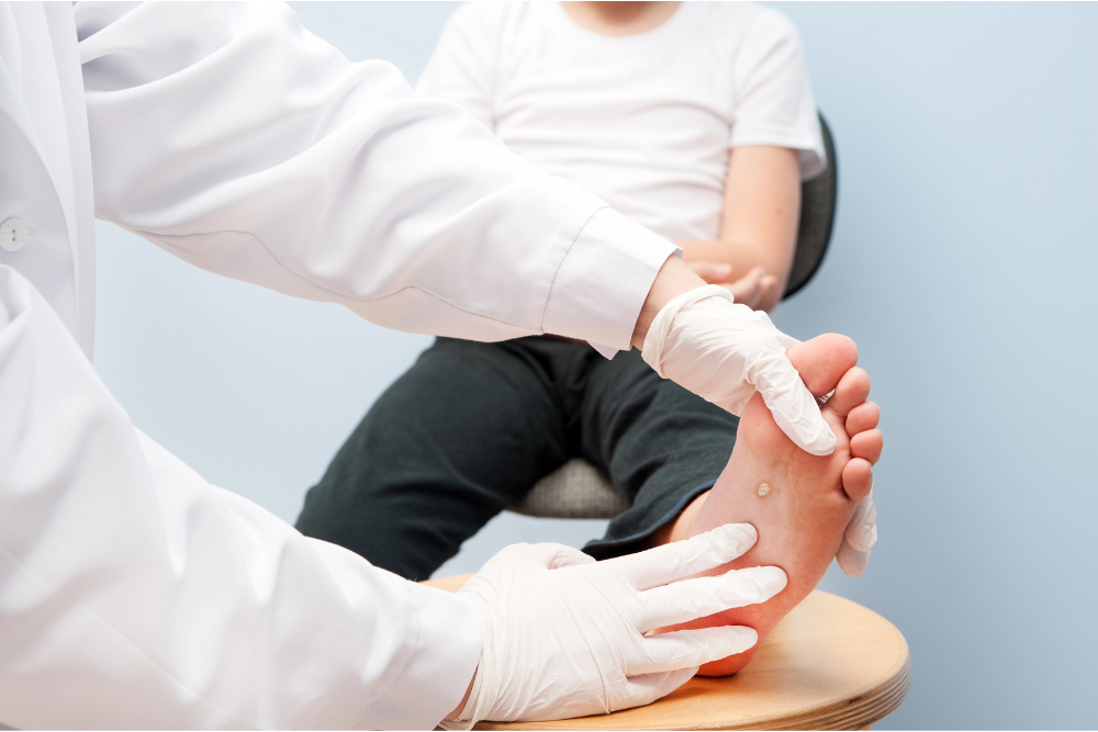
A clinical examination by a podiatrist or vascular specialist is the first step, involving visual inspection of the wound, assessment of pain, and palpation of pedal pulses.
The cornerstone diagnostic tool is the Ankle-Brachial Index (ABI). This non-invasive test compares the blood pressure measured at the ankle to the blood pressure measured at the arm:
● An ABI of 0.90 to 1.30 is normal.
● An ABI above 1.30 suggests vessel calcification (common in diabetes).
● An ABI between 0.50 and 0.90suggests mild-to-moderate PAD (often compatible with VFU treatment).
● An ABI below 0.50 indicates severe PAD and is diagnostic of an arterial ulcer.
Doppler ultrasound and other vascular imaging studies are frequently employed to confirm the extent of arterial blockage or venous reflux, providing the surgeon or wound care specialist with the necessary road map for intervention.
Early and accurate diagnosis prevents complications, including unnecessary tissue loss and infection.
Treatment for a venous ulcer is focused on reversing the underlying venous hypertension to get the blood flowing back toward the heart.
The single most important intervention is compression therapy using specialized multi-layer bandages or prescription-strength compression stockings to apply gradient pressure to the leg. This external pressure counteracts the high internal venous pressure, preventing fluid leakage and aiding venous return.
Other critical elements include:
● Elevation and Exercise
● Advanced Wound Dressings
● Addressing Underlying Disease
Treatment for an arterial ulcer is entirely different and centers on restoring adequate blood flow, as the wound will not heal until the oxygen supply is corrected.
Compression therapy is strictly contraindicated if the ABI is low or if severe PAD is confirmed, as it would further restrict the minimal blood flow, leading to rapid tissue death.
Treatments include:
● Revascularization
● Pain Management
● Systemic Control
● Specialized Wound Care
In cases where conventional treatments stall ,advanced wound care strategies are employed for both ulcer types under coordinated care. The include:
● Debridement: Removing dead tissue is necessary for healing, often using surgical, enzymatic, or autolytic methods.
● Biologics: Skin grafts, bio-engineered skin substitutes, or application of growth factors can accelerate healing in difficult wounds.
● Negative Pressure Wound Therapy (NPWT): A vacuum dressing system that pulls exudate away, reduces edema, and increases blood flow to the area.
● Hyperbaric Oxygen Therapy (HBOT): For selected arterial ulcers, HBOT may be used, where the patient breathes 100% oxygen in a pressurized chamber, saturating the blood plasma with oxygen to help damaged tissue survive.
Patients at high risk, particularly those with diabetes, hypertension, or a history of PAD, must engage in rigorous long-term management.
Daily self-inspection of the feet is essential, and high-risk patients should receive regular, professional foot examinations from a podiatrist.
Additionally, custom-fitted orthotics or orthopedic footwear help prevent the pressure points that lead to arterial ulcers.
Strict control of blood sugar, blood pressure, and lipid profiles is the most effective preventative measure against the vascular damage that causes both ulcer types.
Finally, lifestyle modifications like smoking cessation (critical for PAD) and consistent exercise help improve overall circulation.
Podiatrists are the first line of defense, providing early diagnosis, conservative wound care, and necessary referrals to vascular specialists. Consult a podiatrist or wound care specialist immediately if you notice:
● An ulcer is not showing signs of improvement after two weeks of basic care.
● Indications of infection include spreading redness, warmth, pus, fever, and foul odor.
● Severe or rapidly worsening pain, especially pain that wakes you at night or requires you to hang your foot off the bed.
● Color changes in the foot or toes ,such as blue or black.
Successful healing of foot ulcers requires a correct diagnosis by a podiatrist as to whether the wound is caused by a failure of blood return (Venous) or a failure of blood supply (Arterial).
This distinction dictates the treatment path: compression for venous insufficiency and revascularization for arterial blockage.
By leveraging specialized testing, such as the ABI, and integrating advanced wound care strategies, patients can achieve effective healing, better outcomes, and a significant reduction in the risk of limb loss.
If you or a loved one is struggling with a chronic foot wound, request an appointment today at Advanced Foot and Ankle Specialists.
